Instrument specification
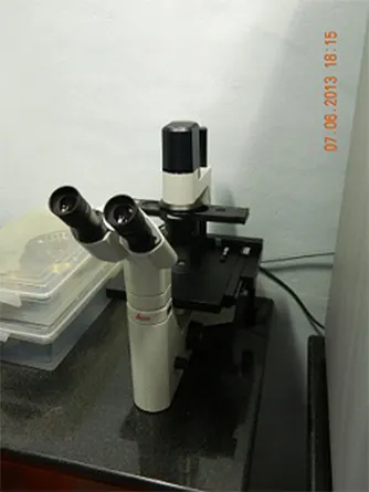
The confocal Raman Microscope, CRM alpha 300 S (WITec GmbH) is the best of its class instrument that combines in a unique way the operations of confocal microscopy, Raman spectroscopy, AFM and SNOM. This work station has been used for: (i) Confocal microRaman spectroscopy and microscopy (Raman spectrum for different varieties of samples such as solids, powders, thin films, liquid and so forth, SERS [air and liquid measurements], Raman spectral imaging), (ii) confocal section analysis, depth scanning in transmission and reflection modes, (iii) atomic force microscopy (contact and tapping modes), and (iv) aperture scanning near field optical microscopy (SNOM).
The confocal setup reduces unwanted background signals, enhances contrast and provides depth information. Differences in chemical composition, although completely invisible in the optical image, will be apparent in the Raman image and can be analyzed with a resolution down to 200 nm. Its sensitive setup allows for the nondestructive imaging of chemical properties without specialized sample preparation. With AFM, investigation of material properties on the nanometer scale is possible. As SNOM requires only minimal sample preparation if any, it is ideally suited to quickly and effortlessly image the optical properties of a sample with resolution below the diffraction limit. Typical applications are found in nanotechnology, materials research, life sciences and others.
Technicalities
Key features
- Spatial resolution beyond the diffraction limit (ca. 60 nm laterally)
- Unique patented SNOM sensors
- Ease of use in air and liquids
- Nondestructive, label-free imaging technique provides super-resolution microscopy with minimal, if any, sample preparation
- Upgradeable with confocal Raman imaging for correlative microscopy and near-field Raman imaging
- Three techniques always integrated within one instrument: confocal microscopy, AFM and SNOM
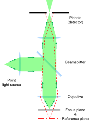
Theory of operation
About the Instrument:
Selected Data:
Footnotes:
1. http://www.witec.de/en/home
2. Using ambient ion beams to write nanostructured patterns for surface enhanced Raman spectroscopy, Anyin Li, Zane Baird, Soumabha Bag, Depanjan Sarkar, Anupama Prabhath, Thalappil Pradeep and R. Graham Cooks, Angew. Chem. Int. Ed., 2014, 53, 12528-12531
3. Single- and few-layer graphene growth on stainless steel substrates by direct thermal chemical vapor deposition, Robin John, Ashok Reddy, C. Vijayan and T. Pradeep, Nanotechnology, 2010, 22, 12528-12531.
4. Ambient electrospray deposition Raman spectroscopy (AESD RS) using soft landed preformed silver nanoparticles for rapid and sensitive analysis, Tripti Ahuja, Atanu Ghosh, Sandip Mondal, Pallab Basuri, Shantha Kumar Jenifer, Pillalamarri Srikrishnarka, Jyoti Sarita Mohanty, Sandeep Bose and Thalappil Pradeep, Analyst, 2019, 144, 7412-7420.
5. Organic solvent-free fabrication of durable and multifunctional superhydrophobic paper from waterborne fluorinated cellulose nanofiber building blocks, Avijit Baidya, Mohd Azhardin Ganayee, Swathy Jakka Ravindran, Kam Chiu Tam, Sarit Das, Robin Ras and Thalappil Pradeep, ACS Nano, 2017, 11, 11091−11099
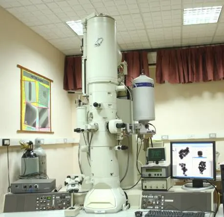
High-resolution transmission electron microscopy (HRTEM)
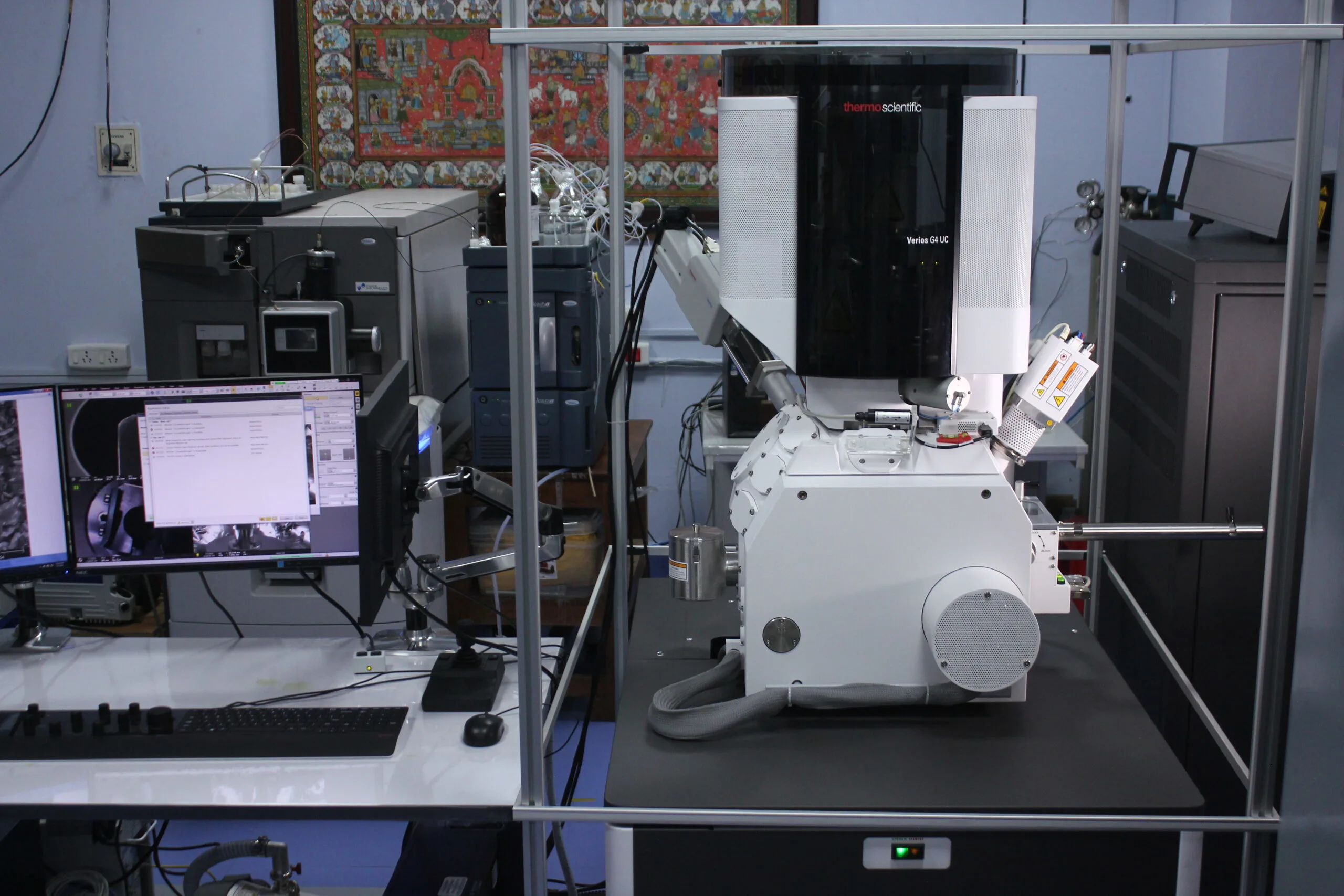
Field Emission Scanning Electron Microscope
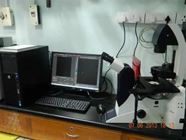
Hyperspectral Imaging
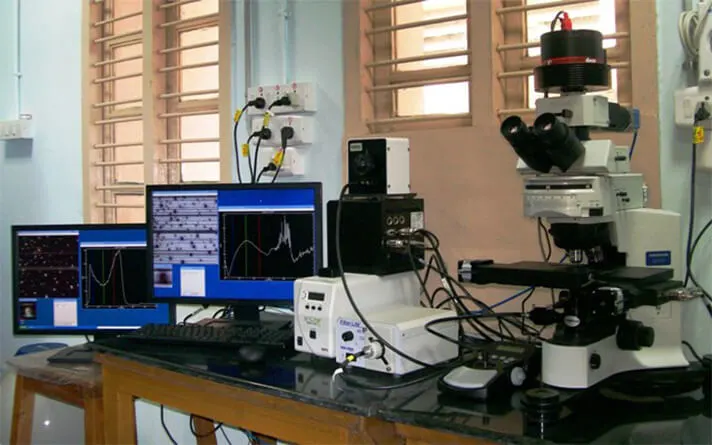
Phase Contrast Microscope
Instrument Specification: Available magnifications: 4X, 10X, 20X and 40X phase